
Researchers with the Department of Neuroscience at the University of Arizona have discovered neurons within the brain that control one’s urge to eat in response to inflammation. On June 24, their findings were published in Nature Communications.
Members of the neuroscience research team and co-authors of the paper, Yong Wang, JungMin Kim, Mathew Schmit, Tiffany Cho, Caohui Fang and Haijiang Cai, Ph.D., investigated the bed nucleus of the stria terminalis’ (BNST), a region of the brain, and it’s connection to feeding behavior.
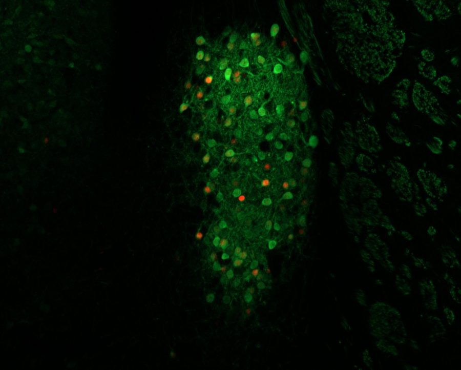
According to the paper, titled “A bed nucleus of stria terminalis microcircuit regulating inflammation-associated modulation of feeding,” in the BNST, a region of the brain closely related to the amygdala, researchers found “a population of neurons” that regulate “general feeding behaviors.”
This cluster of neurons may provide a new target for treating anorexia and other eating disorders, said Cai, an assistant professor at the UA and BIO5 fellow who heads the lab that made the finding.
“A mid-term goal of my lab … is if we can tease out the neural circuitry that control feeding and the neural circuitry that control anxiety,” Cai said. “Hopefully in the future we can design drugs for … eating disorder patients.”
Schmit, a UA graduate student of neuroscience, noted the importance of these neurons to more targeted treatments that control feeding without side effects.
“People don’t eat for reasons that are usually unpleasant,” Schmit said, citing nausea, sickness and undesirable food. “Let’s say that we want to stop feeding but not in an unpleasant way … These cells, when you turn them on, you don’t feel [worse].”
In other words, though the BNST is associated with feeding as it relates to inflammation, which usually makes one feel ill, activating or deactivating those neurons does not have an effect on the way one feels.
The research team experimented on mice to reach their conclusion. Using a method known as optogenetics, researchers injected a virus into mice’s brains that allowed them to activate a neuron at will using light, Schmit said.
“What we’ve done is we’ve hijacked a protein from a bacteria,” Schmit said. “What it does, basically, is every time light hits it, it pops open and lets a bunch of positive charges into the neuron and that makes it fire. That means we can shine a light on a neuron … and make it fire. We can literally control neurons with light.”
Schmit wrote the behavioral analysis program that allowed researchers to monitor mice’s eating behaviors while the neurons were active or inactive.
“Basically … we videotape the mouse in its cage and … look at things like how long it takes the mouse to approach food … [or] measure how long it’s eating for or … to know if [the mouse] is sated,” Schmit said.
Research technician Cho, who, at the time of the study, was working on her bachelor’s in neuroscience and cognitive science, helped analyze the mice’s behavior.
“I … watched videos of mice and … recorded time points of when they were eating and when they weren’t,” Cho said.
The research team concluded that when the group of neurons in the BNST were activated, mice would stop eating. Likewise, when they were deactivated, the mice would eat.
“Neurons in this brain region called BNST … are activated during this inflammatory response [that suppresses feeding behavior], and so this type of neuron is interesting because when we silence these neurons … then we can … block the feeding suppression,” Cai said.
In 2014, Cai identified a region within the central amygdala, the brain’s emotional hub, that controlled a portion of feeding behaviors.
“Because the BNST is so closely connected with the central amygdala … which is Dr. Cai’s post-doc work, we’re just looking for whatever comes out of the amygdala,” Cho said. “We’re kind of seeing what offshoots from that. So, kind of everything is pretty much stemming from Dr. Cai’s 2014 paper.”
Combined with the recent discovery in the BNST, the department of neuroscience at the UA endeavors to better understand the brain’s full process behind feeding behaviors in future research, Schmit said.
“We like to tell a nice, neat story when we write a paper, but really research is a chain of events,” Schmit said. “A paper is just a snapshot of events that might go further.”
Follow Sam on Twitter