Every passing minute is crucial as a small team of students attempt to revive their patient, who lays lifeless on the table before them. A team leader calls out directions, while several students perform essential tasks, such as exhausting chest compressions and providing ventilation.
Finally, a pulse appears on the screen, letting the team know the procedure was a success.
“He’s alive!” one student shouts. The team claps and congratulates one another, but only for a few short moments before jumping back into a simulation room for the next scenario. This particular simulation is called an Interprofessional Education and Practice Cardiopulmonary Resuscitation Team Behavior Simulation and includes medical, pharmacy and nursing students.
While the adrenaline and challenges faced are real, the emergency situation is not. It is just one of the many simulations performed at the Arizona Simulation Technology and Education Center (ASTEC) at the University of Arizona College of Medicine.
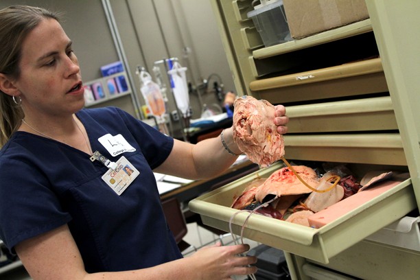
The lab was started in August 2005 and Allan Hamilton, executive director of the lab, has been designing tissue since the early 90s — beginning with tissue for neurosurgery. Since designating a space for tissue design when the lab opened in 2005, the lab has expanded its materials to what it uses today.
The lab, like most simulation labs, uses high-fidelity mannequins to practice emergency medicine for real life situations. High-fidelity mannequins can breathe and have a pulse, according to Lisa Grisham, a medical simulation specialist at ASTEC.
However, ASTEC is one of the few labs to have an in-house tissue lab that can create and build actual training models for surgical procedures, according to David Biffar, director of operations at ASTEC and a creator of the simulated tissues used.
Together, this technology creates a realistic simulation lab complete with mannequins that can breathe, have a pulse, blink their eyes and even bleed.
The mannequin is hooked up to and controlled by a computer. Simulation specialists can control the mannequins’ symptoms, leaving the students to respond as if it were a real emergency.
They can make its heart and lungs sound abnormal, the electrocardiogram tracing abnormalities and certain parts of the body unable to function properly — essentially making it look the same as if a real person was having those problems, according to Grisham.
“Right now, it’s about the closest we can get without using a real patient,” Grisham said. “The benefit of this is we don’t have to use animals, we don’t have to go to the cadaver for tissues … we can do a lot of the same things using this, but it’s not perfect.”
Specialists are also able to place a small microphone in the mannequin’s mouth so that students can talk with the patient, though they don’t take it to that level, Grisham said.
“We want them [students] to really interact with the mannequin, and check his pulses, and make sure that he has a pulse,” Grisham said.
The tissue lab has created tissues to allow students to practice drawing blood and inserting IVs, as well as simulate some injuries. A tube that circulates synthetic blood can be run through the tissue of the mannequin, and up through an open wound to make it bleed. Students then have to address the wound and wrap it up so the patient does not bleed out, according to Grisham.
Biffar said he spends about two hours a week creating tissues that are either not available on the market or need to be upgraded to address a limitation it has.
“It opens up the gates for opportunities to be a part of grants, or to pursue grants. It’s kind of nice to have this and not many simulations do,” Biffar said.
The lab uses casting and molding techniques and materials such as ballistic gelatin, silicone and hot vinyl. Biffar can create tissues such as suture pads, anastomosis models — a practice model typically used by neurosurgeons to connect one vessel to another — fractures and abrasions.
He creates these tissues based on how they might be used for multiple attempts and with the best material to maximize its utilization, he said.
“Simulation is not going anywhere anytime soon … you can spend eight hours on making something super realistic and you can only use it once,” Biffar said. “Or you can find a way to kind of flesh out the most important part of a procedure, and try to make it so you can replicate it.”
Kevin Severson, a finance and physiology senior, ASTEC volunteer and CPR instructor, said he enjoys seeing how the students learn from their mistakes, as well as interacting with students in general.
“Aside from that, being in this lab, obviously I got to learn how to … insert IVs, draw blood, train on laparoscopic trainers, work with a research team here a little bit, so it’s helped me to get a broader view on the medical arena,” Severson said.


