
University of Arizona professors and students are collaborating to take exponential steps toward finding a cure to ovarian cancer.
Jennifer Barton is the brains behind the whole operation, and her new device, the “falloposcope,” has plans to change modern medicine around diagnosing ovarian cancer. Barton is a biomedical engineering professor at the university and also directs the Bio5 institute on campus.
When Barton first came to the university, she met with a physician who requested that she try to create something to diagnose patients in the early stages of ovarian cancer.
“When a physician comes to a biomedical engineer and says ‘I need you to build something to solve this horrible problem,’ then engineers always go, ‘yes, okay, I’ll take that challenge on,’” Barton said.
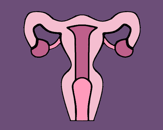
According to Barton, ovarian cancer starts in the fallopian tubes — tubes that transport the egg from the ovaries to the uterus. Much like cervical cancer, it develops from a few abnormal cells. The cells start proliferating and they can stay there for a number of years, yet everything appears fine to the host.
“If we could catch the cancer in that 6-year window, then we can cure the disease with surgery or chemotherapy,” Barton said.
However, a lack of modern technology once prohibited oncologists from getting inside the fallopian tubes to do screenings for ovarian cancer.
“Once those cells leave the fallopian tubes, they float out and then attach themselves to the ovaries or other places in the abdominal cavity and they, for reasons we don’t entirely understand, will start growing very quickly,” Barton said. “It’s not until they have grown very quickly that you start having symptoms and the disease has advanced.”
Most people who exhibit these symptoms are then diagnosed with ovarian cancer. Because they usually are in a very late stage of their prognosis, their outcomes are typically very poor. People with stage four ovarian cancer have a relative five-year survival rate of 17%.
“For a long time people didn’t really know why, because for other cancers, like cervical cancer, you have pap smears every year,” Barton said. “Generally in the United States, you catch cervical cancer very early. But, there’s no screening like that for ovarian cancer.”
Barton realized that to catch cancer early, doctors have to get something inside of the fallopian tubes. The cancer cells in the fallopian tubes are too small to be detected by CT, MRI, ultrasound or any comparable sort of imaging modality.
“You really need to use optics; you have to look and analyze it,” Barton said. “The problem with optics is we can’t look into our body.”
Barton and her team have figured out a mechanism in which they can go through the uterus and into the fallopian tubes.
“It’s just a little bit of a challenge to make something small enough to do that,” Barton said.
Challenge aside, her team was able to make the “falloposcope” to go into the fallopian tubes, shine light on the tissue, collect the light that comes back, analyze it and determine whether or not cancer is present.
“Technology is getting to the point where we have these little tiny fibers and these miniature sensors and we have these new materials that are flexible and pushable and biocompatible,” Barton said.
In the past, cancer biologists have taken tissue that was being removed in surgery anyway and looked at it in the laboratory. This way, they were able to make sure that the optical techniques will allow them to detect cancer. The next step is seeing if they can get their microscope into the body rather than bringing the tissue to the microscope.
Dr. John Heusinkveld works with Barton in his own personal OB-GYN practices here at the UA. Heusinkveld is a UA alumni who now practices at Banner. He has assisted in the trial of the falloposcope on patients, which is the first step in the development of the device.
The current stage of the study consists of rounds of surgery on people who are not at risk for cancer and do not currently have cancer. Heusinkveld is not a cancer surgeon, but he does complete surgeries on women who are having their fallopian tubes removed for various reasons. The people who Heusinkveld operates on are volunteers who have agreed to extend their surgery so that doctors can try out the cameras and make sure the mechanics are up to par.
“My role is that I ask people if they are interested in participating and if they do agree in participating, I am the one who looks at the fallopian tubes with the device before I remove them,” Heusinkveld said. “Because we’re moving toward something that will eventually help with cancer care, a lot of women are willing to do this. We’re not going to extend their anesthesia time for more than 15 minutes and there’s no harm to these patients.”
In this trial, the main priorities are to find out what a normal fallopian tube looks like and ensure that the operation does not cause harm to the fallopian tubes.
“There is a huge variation between people,” Barton said. “You really have to understand what normal looks like before you can determine what disease looks like.”
The next step after this study is to test the falloposcope in people that are at high risk for ovarian cancer.
Barton has been working on this study for over 20 years, but may be nearing the finalization of her device if the next stages of development run smoothly. Barton’s said her goal is to get a medical device company to license this technology and start producing it once it has completed sufficient trials.
“My students and I are building like 30 or 40 of these endoscopes in the basement of our building and we can do that, but we can’t build hundreds or thousands,” Barton said.
Andrew Rocha is a Ph.D. candidate in the optical science program at the university who is working alongside Barton. Rocha needed to find a new project two years into his Ph.D. and a new principal investigator who had funding. Barton had made a callout to the college, saying that she had a funded grant to do some endoscope work.
“When I applied with her, I let her know that most of my experience was building space optics and most of my internship experience had to do with laser communication satellites and optical systems,” Rocha said.
Although out of his original field of interest, Rocha knew that this was a notable opportunity to try something new. Initially, he started on his first project which was making an endoscope with the application of trying to do early detection of pancreatic cancer.
“I wanted to absorb as much information as I could, so I hopped onto the falloposcope project because they were in the middle of building endoscopes. I had never built anything that small before; I wanted to get that exposure,” Rocha said.
Rocha was able to continue on this team and took part in the initial trials with Heusinkveld. He helped continue the pilot study, which is making sure that the endoscopes are meeting all of the specifications of the design.
Before this, Rocha mentioned that he had never even set foot in a surgery room, let alone instructed a doctor on how to use their device.
“I think the challenging part about it is we are essentially doing things that have never been done before so everything is a learning lesson, … learning how our medical device is going to be used versus how it works in the lab,” Rocha said. “Everything is so different and I think that’s a really tough part of it — trying to anticipate how things are going to happen in the real world.”
Follow Annabel Lecky on Twitter