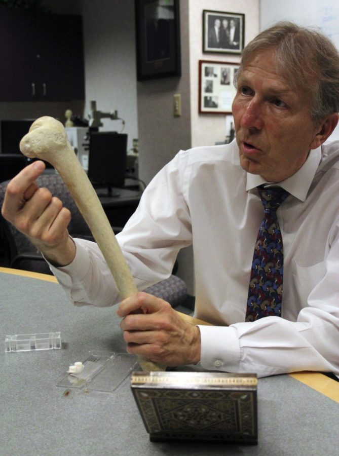
New research on tissue regeneration between the UA Orthopaedic Research Laboratory and the Arizona Arthritis Center might offer a new alternative to joint replacements.
The most common type of arthritis is called osteoarthritis, where the cartilage in the joints begins to break down, causing it to stiffen and lose its elasticity, according to John Szivek, the laboratory’s director.
Instead of using artificial joints to repair the damage, which last 25 to 30 years with regular replacements, tissue regeneration procedures would re-grow existing tissue.
The procedure begins by scanning the bone material of a joint with a high-resolution computed tomography scanner, a machine that uses X-rays to create detailed 3D images of the inner parts of the body. This machine then sends the exact material information to a 3D printer that will construct a small scaffold with the exact structure of the bone to act as carrier for the cartilage to grow upon.
“These are for what is typically called Focal Defect Repair Systems, so if there’s a focal defect or damaged spot, you can use these to repair that spot,” Szivek said.
Scientists would then drop cells on that scaffold to grow the surface of the cartilage. However, making sure the tissue is the right kind of tissue is the challenge, according to Szivek.
“The cells (in cartilage) are highly structured in specific places and build the tissue that way,” Szivek said. “So one of the things we’ve been looking at is how we can align the cells in a specific pattern.”
Szivek discovered that by adding a container to the scaffold, they could align the strands of the cells in one direction using the adult stem cells found in fat tissue.
“The nice thing about adult stem cells is there are lots of them found in fat, they’re easy to harvest, you can collect them pretty easily, they don’t mind growing in flat culture and we can convert them to cartilage cells reasonably easy,” Szivek said. “If we’re happy to wait, we can just drop them into the cartilage itself and all the other cartilage cells will convince them to become cartilage cells.”
According to Szivek, understanding how and why those tissues grow the way they do is very useful when comprehending their regenerative response. When participating in activities like walking, running and jumping, stress is exerted on the tissue. The information of the work load is crucial so that the patient ends up with the correct type of tissue.
Szivek’s team has been using tiny strain gauges that attach to a radio transmitter to determine the load bearings on joints.
“We’ve tried placing the transmitter in dogs, and depending on how much they load it, the strain gauges tell us how much they are loading their knee,” said Andrea Arellano, an undergraduate student in biomedical engineering. “They send that information through the transmitter using a power coil. The transmitter then gets the data and sends it to our handheld system so we can analyze that data.”
The study is conducting experiments on other parts of the body as well. Testing is still in its early phases, so most of the experiments are being conducted on animals. The lab is provided with some discarded tissues from patients but the materials become costly.
“In my experiment, we seed these bone plugs and let them grow for 25 days and at the end we plan on doing several different methods to see if the gene expression changes at the end,” said Marysol Luna, an undergraduate student in biomedical engineering.
“We’re at the start of the process of collecting the information the FDA would need to allow us to now use these processes on a patient,” Szivek said. “How long then it will take us to get something through to the FDA is anybody’s guess.”


