Apple earbuds are now wireless and if a new UA research project on acoustoelectric brain imaging goes well, brain scans could be next.
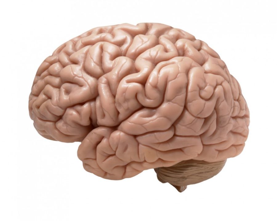
ABI technology uses sound waves to measure the amount of electrical activity in the brain and produces a high-resolution image—potentially less than 5 millimeters of specific resolution—in real time. This noninvasive technique works faster than current technologies.
Dr. Russell Witte, a UA assistant professor of radiology, optical sciences and biomedical engineering, is the principle investigator for the project. He previously worked with acoustoelectric cardiac imaging, but said his “interest is ultimately very focused on the brain.”
RELATED: Parker Antin takes science seriously
The three-year project started in October of 2015 with a $1.15 million National Institutes of Health grant. The project will work on measuring current source density or where currents sink and source in the brain and exploring the preferred pathways between regions in the hippocampal network.
This technology could be used to help diagnose behavioral and neurological diseases, like addiction, epilepsy and Parkinson’s, as well as help doctors more precisely prescribe the right amount of medication, according to Witte.
Witte said this first year has primarily been about developing the ultrasound instrumentation, which is no easy task considering that most need to be custom-made and programmed.
“A lot of the instruments we can’t just get off a shelf,” Witte said. “We have to do a lot of bench testing first to get proof of concept and to make sure there’s no risk.”
Two of the main challenges Witte and his team have run into are the small size of the signals they’re planning on studying and the way the skull blocks the sound waves.
“We’re getting into an area where we don’t really know much about the mechanics of the brain,” Witte said.
The UA is collaborating with the University of Washington for ultrasound and probe engineering, the University of Michigan for skull density issues and Duke University, with neurosurgeons.
“The idea is we all kind of learn about the issues,” Witte said. “We have a lot of different expertise working together.”
But, not all of that expertise comes from outside UA. Neuroscientist Stephen Cowen, a UA assistant professor of psychology, has been busy setting up for the second phase of the project: testing the technique on rats.
Cowen’s “current success” is measuring what a baseline brain wave, or volley, looks like in rats.
“We’ve been finding a way to trigger a reliable brain wave by putting an array of electrodes in the hippocampus, then triggering it to send a burst of activity,” Cowen said.
RELATED: Neuroscience students and faculty showcase research at Data Blitz
Cowen has more than 15 years of experience working with electrodes, and again, size is the biggest obstacle. Cowen needs to “miniaturize” all of the instruments, both to work with the animals and to not interfere with the ABI equipment.
“Ultrasound machines are usually used on people, but I deal with rats,” Cowen said. “It’s a challenge to adapt the system. Rats are great because we know how their brains are set up … but they’re tiny.”
Physiological sciences graduate students Daniel Hill and Cameron Wilhite are at the forefront of the process to make the electrode arrays smaller. Both joined this project over the summer, though Hill said his main role was helping Wilhite build and implant the arrays.
Wilhite spends between four and five hours on each array, which is composed mainly of two bundles of four wires that are thinner than a human hair. Those wires pick up on volleys and track how they change “downstream activity” in the brain.
Witte said that ABI could have a “broad impact,” especially if it can be made portable, similar to EEG systems used with iPhones.
“Having something portable and accessible in real time would benefit many people,” Witte said. “To have another tool that could be utilized by a lot of different people could redefine research.”
Follow Marissa Heffernan on Twitter.

